SVT vs. Other Heart Conditions Quiz
Test your knowledge of how to distinguish Supraventricular Tachycardia (SVT) from similar heart conditions. Select the correct answer for each question.
Question 1: What is the typical heart rate range for SVT?
Question 2: Which condition has an irregularly irregular pulse?
Question 3: What ECG finding distinguishes Ventricular Tachycardia from SVT?
Question 4: What typically triggers Sinus Tachycardia?
Comparison Table: Key Features
| Condition | Heart Rate | Pulse Regularity | ECG Findings | Typical Trigger |
|---|---|---|---|---|
| Supraventricular Tachycardia | 150-250 bpm | Regular | Narrow QRS | Caffeine, stress |
| Atrial Fibrillation | 100-180 bpm | Irregularly Irregular | Irregular rhythm, no P waves | Alcohol, sleep apnea |
| Atrial Flutter | 150-300 bpm | Regular (often 2:1 block) | Flutter waves | Heart disease, thyroid issues |
| Ventricular Tachycardia | 150-250 bpm | Regular | Wide QRS (>120 ms) | Heart disease, electrolyte imbalance |
| Sinus Tachycardia | 100-150 bpm | Regular | Upright P waves | Exercise, fever, anxiety |
When your heart starts racing unexpectedly, it’s natural to wonder whether you’re dealing with a harmless flutter or something more serious. Supraventricular Tachycardia (SVT) is a common type of rapid heartbeat that originates above the heart’s ventricles, but it often gets confused with other arrhythmias or heart issues. This guide walks you through the key characteristics, diagnostic clues, and treatment options that set SVT apart from its look‑alikes, so you can recognize the signs and seek the right care.
Key Takeaways
- SVT arises from electrical impulses above the ventricles and typically produces a regular rate of 150‑250bpm.
- Symptoms overlap with atrial fibrillation, atrial flutter, and sinus tachycardia, but ECG patterns and onset triggers differ.
- Quick bedside tests (pulse check, symptom timing) and a 12‑lead ECG are the fastest ways to tell them apart.
- Management ranges from simple vagal maneuades to medication or catheter ablation, depending on frequency and severity.
- Keeping a symptom diary and sharing it with a clinician speeds up diagnosis and treatment planning.
What Is Supraventricular Tachycardia?
Supraventricular Tachycardia is a rapid heart rhythm that starts in the atria or the atrioventricular (AV) node, the area just above the ventricles. It’s characterized by a sudden onset, usually lasting seconds to hours, and a heart rate that often exceeds 150 beats per minute. The condition can affect anyone, but it’s most common in people aged 15‑40 and in those with a family history of arrhythmias. Because the electrical circuit stays above the ventricles, the ventricular contraction remains coordinated, which is why patients often feel a “flutter” rather than a full‑blown collapse.
Common Heart Conditions That Mimic SVT
Several other arrhythmias and cardiac issues can look like SVT on first glance. Below are the most frequently mistaken conditions, each introduced with its own microdata definition.
Atrial Fibrillation is an irregular, often rapid heart rhythm originating in the atria, characterized by chaotic electrical activity and an irregularly irregular pulse.
Atrial Flutter is a fast, regular rhythm that typically originates in a re‑entrant circuit around the tricuspid valve, producing a “saw‑tooth” pattern on ECG.
Ventricular Tachycardia is a life‑threatening rapid rhythm that begins in the ventricles, often presenting with a wide QRS complex and a risk of progressing to ventricular fibrillation.
Sinus Tachycardia is an appropriate increase in heart rate (usually < 100bpm) that originates from the sinus node, often in response to exercise, fever, or anxiety.
Premature Atrial Contraction is an early beat that originates in the atria, usually harmless, but can feel like a skipped beat or brief flutter.
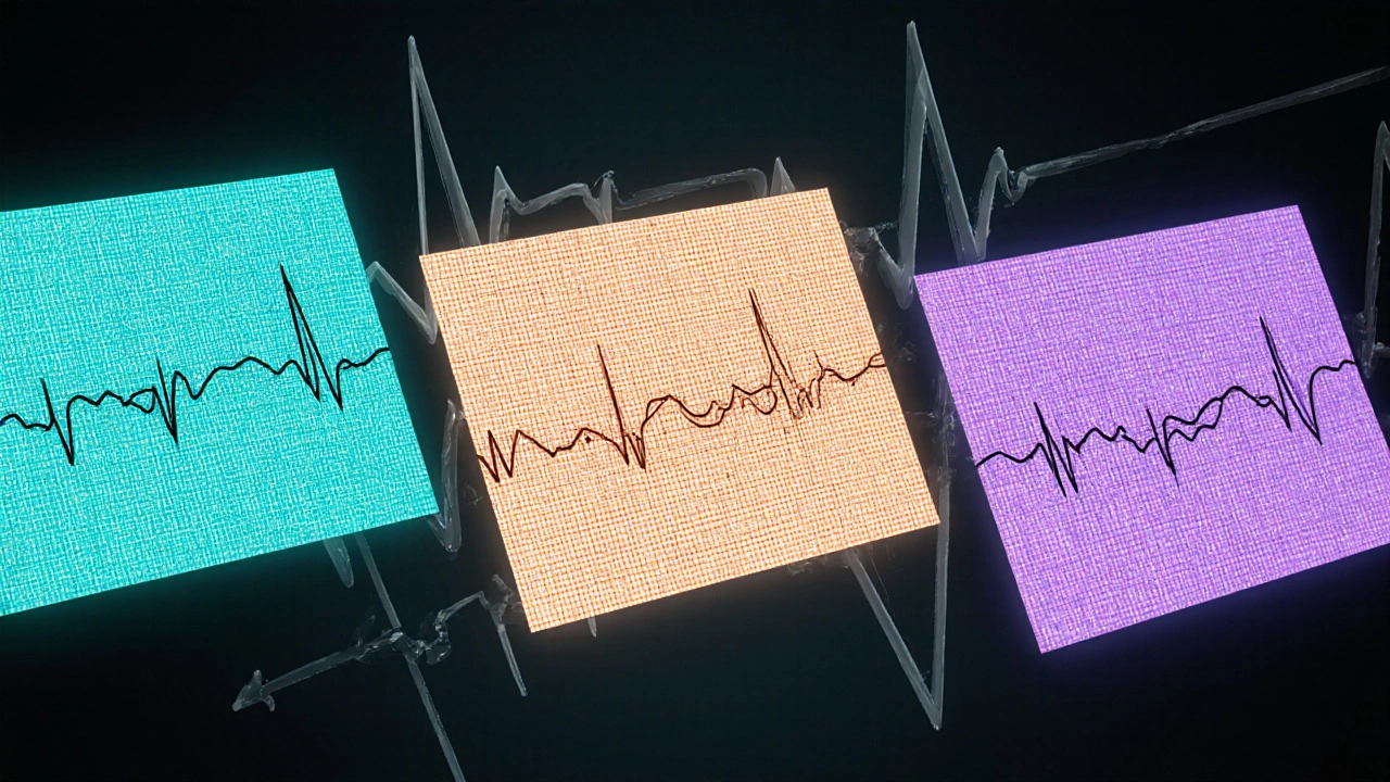
Electrocardiogram (ECG) is a non‑invasive test that records the electrical activity of the heart, providing the primary visual cue for distinguishing arrhythmias.
Electrophysiology Study is an invasive diagnostic procedure where catheters map the heart’s electrical pathways to locate the precise origin of an arrhythmia.

How to Spot the Differences: Symptoms and Triggers
While many arrhythmias share palpitations, dizziness, or shortness of breath, certain patterns help narrow the culprit.
- Onset speed: SVT usually starts abruptly, often after a caffeine boost, stress, or a sudden change in posture.
- Pulse regularity: SVT produces a regular, fast pulse; atrial fibrillation feels irregular; atrial flutter feels regular but at a slightly slower rate (150‑250bpm with a 2:1 block).
- Duration: SVT episodes may self‑terminate within minutes, whereas atrial fibrillation can persist for hours or days without medical intervention.
- Associated symptoms: Light‑headedness or fainting is more common in ventricular tachycardia, while mild fatigue is typical for sinus tachycardia.
- Trigger history: Fever, anemia, hyperthyroidism, or excessive exercise point toward sinus tachycardia; alcohol bingeing or sleep apnea can precipitate atrial fibrillation.
ECG and Diagnostic Tools
Because the ECG is the gold standard, understanding the visual cues can demystify the diagnostic process.
ECG provides a graphical representation of the heart’s electrical activity across four leads, allowing clinicians to measure rate, rhythm, and wave morphology.
Key ECG differences:
- SVT: Narrow QRS (<120ms), regular rhythm, often no visible P‑waves or hidden within the T‑wave (P‑wave‑minus).
- Atrial Fibrillation: Irregularly irregular R‑R intervals, absent distinct P‑waves, presence of fibrillatory waves.
- Atrial Flutter: “Saw‑tooth” flutter waves, usually 2:1 AV block leading to a ventricular rate of ~150bpm.
- Ventricular Tachycardia: Wide QRS (>120ms), regular rhythm, may show AV dissociation.
- Sinus Tachycardia: Upright P‑wave before each QRS, normal axis, rate <100bpm at rest but rises proportionally with activity.
Side‑by‑Side Comparison
| Condition | Typical Rate (bpm) | Origin | ECG Hallmark | Common Symptoms |
|---|---|---|---|---|
| Supraventricular Tachycardia | 150‑250 | Atria / AV node | Narrow QRS, regular, hidden P‑wave | Palpitations, mild dizziness, shortness of breath |
| Atrial Fibrillation | 100‑180 (irregular) | Atria | Irregular R‑R, absent P‑wave, fibrillatory waves | Irregular pulse, fatigue, chest discomfort |
| Atrial Flutter | ~150 (regular) | Atria (re‑entrant circuit) | Saw‑tooth flutter waves, often 2:1 block | Palpitations, shortness of breath, light‑headedness |
| Ventricular Tachycardia | 150‑300 (regular) | Ventricles | Wide QRS, AV dissociation | Syncope, chest pain, sudden collapse |
| Sinus Tachycardia | 90‑180 (regular, activity‑related) | Sinus node | Upright P‑wave before each QRS, normal axis | Usually asymptomatic; may feel “racing” during fever or stress |

Treatment Pathways
Once an accurate diagnosis is made, treatment follows a stepwise approach.
- Vagal maneuvers: Techniques like the Valsalva maneuver or cold‑water face immersion can reset the AV node and terminate an SVT episode.
- Pharmacologic options: Beta‑blockers, calcium‑channel blockers (e.g., verapamil), or anti‑arrhythmic pills (e.g., flecainide) are prescribed based on frequency and comorbidities.
- Cardioversion: A short electrical shock in the emergency department can quickly restore normal rhythm for persistent SVT.
- Catheter ablation: An electrophysiology study identifies the precise pathway, and radiofrequency energy destroys the small tissue area causing the rapid circuit. Success rates exceed 95% for typical AV‑node re‑entrant tachycardia.
- Lifestyle tweaks: Reducing caffeine, managing stress, and treating sleep apnea lower episode frequency.
Quick Checklist for Patients
- Note the exact time an episode starts and ends.
- Record heart rate (use a smartwatch or manual pulse).
- Identify any triggers (caffeine, alcohol, stress, dehydration).
- Bring a recent ECG or Holter monitor printout to every appointment.
- Ask your doctor about a trial of vagal maneuvers before medication.
- Discuss whether an electrophysiology study and possible ablation are appropriate for you.
Frequently Asked Questions
Can SVT be life‑threatening?
Most SVT episodes are benign and resolve on their own or with simple maneuvers. However, very rapid rates can reduce cardiac output, and rare cases may lead to fainting or trigger heart failure in people with pre‑existing disease.
How is SVT different from atrial fibrillation?
SVT produces a regular, fast rhythm that starts abruptly, while atrial fibrillation shows an irregularly irregular pattern with no distinct P‑waves. Treatment strategies also differ: SVT often responds to vagal tricks or catheter ablation, whereas atrial fibrillation may require anticoagulation and rhythm‑control drugs.
What tests confirm an SVT diagnosis?
A 12‑lead ECG taken during an episode is the gold standard. If episodes are sporadic, a Holter monitor (24‑48h) or an event recorder can capture the rhythm. In ambiguous cases, an electrophysiology study pinpoints the exact pathway.
Are there permanent cures for SVT?
Catheter ablation offers a cure in >95% of typical AV‑node re‑entrant tachycardia cases, with a low complication rate. Lifestyle adjustments can also dramatically cut episode frequency.
When should I seek emergency care for a fast heartbeat?
If the rapid rhythm lasts more than a few minutes, is accompanied by chest pain, severe shortness of breath, fainting, or if you have known heart disease, call emergency services. Prompt cardioversion can prevent complications.
Write a comment
Your email address will not be published.


11 Comments
I get how confusing it can be when your heart feels like a drum solo that never stops. The sudden surge of 150 to 250 beats per minute can make anyone panic. It is important to remember that SVT usually starts abruptly and can end just as fast. The regular rhythm is a clue that the ventricles are still working in sync. A quick pulse check can reveal that the beat feels steady rather than irregular. If you can, grab a smartwatch or a phone app and note the rate. Then call your doctor and describe the onset, any triggers like caffeine or stress, and how long it lasted. Most patients find that learning these details helps the clinician choose the right test. Even though the sensation is scary, the underlying electricity is often benign. Keeping a diary of episodes will make future visits much smoother.
Honestly, if you’re still mixing up SVT with atrial fibrillation after reading a basic guide, you’re doing it wrong; the irregularly irregular pulse of AF is as obvious as a broken metronome. This guide lays out the differences like a neon sign – narrow QRS, regular rhythm, and that trademark 150‑250 bpm sprint – so stop guessing and start paying attention.
Hey folks, let’s clear the fog together – SVT is like that friend who shows up uninvited but leaves as quickly as they arrived, while other arrhythmias tend to linger longer. The key is the ECG: look for a narrow QRS complex and a regular rhythm, which points straight to SVT. If you see a wide QRS, you’re probably dealing with a ventricular issue that needs urgent attention. Remember to check the pulse regularity; a consistent beat rules out atrial fibrillation’s chaotic pattern. Also, note any triggers – caffeine, stress, or a sudden change in posture can set off SVT. By sharing this checklist with everyone, we can all spot the signs faster and get the right care without the drama.
Imagine your heart as a drum circle where everyone follows the same beat; when SVT takes over, it’s like a soloist who suddenly speeds up, but the underlying groove stays intact. This phenomenon reminds us that even the most frantic moments have a rhythm we can learn to read. The regular, rapid pulse is a clue that the ventricles are still synchronized, unlike the erratic dance of atrial fibrillation. By practicing a simple breath‑hold or Valsalva maneuver, you can sometimes calm that soloist back into the group. Think of it as a meditation on your own physiology – a moment to observe, not panic. Keep a log of what you ate, how you felt, and any stressors; patterns often emerge. Sharing these observations with your doctor turns a vague complaint into a precise story they can act on. Over time, you’ll gain confidence that you’re not at the mercy of an unknown force, but a partner in the rhythm of life.
Wow, another user claiming they “don’t get” SVT? It’s like watching someone try to decode an ancient language with a children’s picture book – absolutely clueless! The heart doesn’t need your half‑baked theories about “flutter vibes” and “mystery beats.” If you can’t tell a regular 180 bpm sprint from an irregular afib mess, maybe stick to watching Netflix instead of spouting medical nonsense. This guide already spelled out the ECG clues in plain English – narrow QRS, regular rhythm – so stop wasting everyone’s time and actually read it.
Alright, let’s dive into the nitty‑gritty because I can’t stand holding back – SVT is the party animal of the cardiac world, showing up at 200 bpm, dancing in a perfectly regular pattern, and then ghosting you just as fast. It’s not some vague “heart weirdness” you feel after a marathon; it’s a defined electrical circuit above the ventricles that loves caffeine and stress like a sugar‑addicted teenager. If you catch a rapid, regular pulse, you’re probably witnessing this party, not a chaotic afib rave. And guess what? A simple vagal maneuver can be the bouncer that kicks it out for good. So next time you feel your heart doing the cha‑cha, remember it’s just a guest, not a trespasser.
While the prevailing consensus lauds the diagnostic clarity offered by electrocardiography, one must consider whether such reliance inadvertently marginalizes alternative clinical insights. The emphasis on a narrow QRS complex as the hallmark of supraventricular tachycardia, though statistically sound, overlooks the nuanced presentations observed in mixed‑type arrhythmias. Moreover, attributing abrupt onset solely to caffeine or stress diminishes the role of intrinsic autonomic variability, a factor often relegated to anecdote. It is also noteworthy that the literature frequently conflates sinus tachycardia with SVT, despite their distinct electrophysiological pathways. The pedagogical approach that lists “150‑250 bpm” as a definitive range may, in fact, mislead clinicians confronted with borderline cases. A broader spectrum, perhaps extending to 140 bpm in younger athletes, warrants inclusion. Additionally, the categorical dismissal of atrial flutter as merely “regular but slower” fails to acknowledge its potential for rapid 2:1 conduction mimicry. The guide’s suggestion to rely on symptom diaries, while commendable, assumes patient compliance that is rarely achieved in practice. One must also question the implication that vagal maneuvers are universally effective, given documented refractory instances. In light of these considerations, a more integrative diagnostic algorithm, incorporating bedside ultrasound and biomarker assessment, could enhance accuracy. Critics may argue that such complexity burdens primary care, yet the cost of misdiagnosis arguably outweighs the incremental effort. Ultimately, the pursuit of diagnostic elegance should not eclipse the imperative for clinical humility. By embracing a pluralistic perspective, we safeguard against the dogmatic pitfalls that have long plagued cardiology. Therefore, I submit that the guide would benefit from a tempered, more expansive discourse. Future revisions should also address patient education strategies to bridge the knowledge gap.
Great job on the quiz, everyone! If you ever feel that sudden racing heart, try a simple Valsalva maneuver – pinch your nose, exhale against a closed airway, and you might just break the episode. It’s amazing how a few seconds of controlled breathing can reset the AV node and bring your rhythm back to normal. Keep a log of what you ate, how stressed you felt, and any caffeine intake; patterns often pop up that help you avoid triggers. And remember, you’re not alone – many of us have been there, feeling the flutter and then learning to manage it. Stay positive and keep sharing your experiences; the community learns together.
That’s a solid summary – nice work! 😊 Make sure to double‑check the QRS width on the ECG; a narrow complex points to SVT, while a wide one suggests ventricular origin. Also, note the regularity of the pulse; irregularly irregular beats are classic for atrial fibrillation. Keep the terminology consistent and you’ll avoid confusion. 👍
Even though the drama was loud, the core truth remains: SVT’s regular rapid rate differentiates it from the chaotic pulse of afib, and acknowledging that helps cut through the noise.
When we talk about arrhythmias, it’s essential to place SVT within the broader spectrum of tachycardias, recognizing both its unique features and its overlap with other conditions. The hallmark of a narrow QRS complex indicates that ventricular conduction is intact, which is a critical clue that the problem lies above the ventricles. This distinguishes SVT from ventricular tachycardia, where the QRS widens due to abnormal ventricular depolarization. Moreover, the regularity of the rhythm offers a visual cue on the ECG, contrasting sharply with the irregularly irregular pattern seen in atrial fibrillation. While patients often describe a “fluttering” sensation, the underlying mechanism is a re‑entrant circuit or an enhanced automaticity within the atria or AV node. Triggers such as caffeine, alcohol, stress, or sudden postural changes can precipitate these episodes, which typically self‑terminate within minutes. Diagnostic work‑up should begin with a thorough history and a 12‑lead ECG captured during an episode, if possible. In cases where the rhythm is intermittent, a Holter monitor or event recorder may be warranted. Management strategies range from acute vagal maneuvers, like the Valsalva maneuver, to pharmacologic agents such as adenosine, beta‑blockers, or calcium channel blockers. For patients with frequent or refractory episodes, catheter ablation offers a curative option with high success rates. Finally, empowering patients with education about symptom recognition and trigger avoidance can significantly reduce episode frequency and improve quality of life.